I have previously proposed the use of
calcium T channel blockers to treat some types of autism. I did suggest that language might be a good target.
I recently received a question from a
reader who read an abstract from a paper presented to the Brain Foundation,
that suggested Ethosuximide can increase speech in autism. She also asked what
the effective dosage might be.
This subject has come up before in
this blog. Ethosuximide is a very specific T channel blocker, commonly used to
treat absence seizures. Some readers of this blog have already trialed it. The
other interesting one is Zonisamide, which blocks T channels but also has other
effects. We have reports that the starting low dose of Zonisamide had some
interesting beneficial effects that were lost at the regular higher doses.
I did not expect to find much new
information, but that changed when I found the patent document submitted by
Charles Niesen. So here is a blog post dedicated to this specific subject.
Here is the full patent:
Method
of treating expressive language deficit in autistic humans
Here is an easy-to-read summary:
A New Patent
Claims an Unusual Approach to Autism Language Deficits
A recent patent proposes a novel
pharmacological method for improving expressive language in individuals with
autism. Rather than introducing a new drug, the invention repurposes a class of
existing anticonvulsant medications—specifically succinimides such as
ethosuximide, methsuximide, and phensuximide.
These drugs have long been used to
treat epilepsy, particularly absence seizures. However, the patent suggests
they may also address one of the most challenging aspects of autism: the
inability to initiate and sustain meaningful verbal communication.
Understanding the
Problem
Autism is often characterized by
difficulties in social interaction, but a core feature—especially in more
severe cases—is expressive language impairment. Many individuals with autism
may speak only in short phrases or single words. Others may respond to
questions but rarely initiate conversation or engage in back-and-forth
dialogue.
This is distinct from related
conditions like Asperger syndrome, where language is typically intact but
social communication is impaired. In classic autism, the issue is not just how
language is used—but whether it emerges spontaneously at all.
Currently, there are no FDA-approved
medications specifically designed to improve expressive language in autism.
Most available treatments focus on associated symptoms such as irritability,
seizures, or attention deficits.
The Core Idea
Behind the Patent
The patent proposes that daily
administration of a succinimide anticonvulsant—most notably ethosuximide—over
an extended period (typically several months) can significantly improve
expressive language abilities.
Patients are treated for at least one
month, with stronger effects reported after three to six months or longer. The
goal is not just increased vocabulary, but a progression toward spontaneous
speech and true conversational ability.
How Might This
Work?
Ethosuximide works by blocking T-type
calcium channels in the brain. These channels play a role in regulating
neuronal activity and rhythmic signaling.
While the exact mechanism in autism is
unknown, the patent speculates that modulating these channels may help
normalize communication between brain regions involved in language. Another
hypothesis is that the drug may “activate” previously underused or dormant
neural circuits.
These ideas remain theoretical and are
not yet confirmed by broader research.
Dosage and
Treatment Approach
The proposed dosing follows standard
epilepsy guidelines, typically ranging from 10 to 60 mg per kilogram of body
weight per day. In many cases, a range of 20–40 mg/kg/day is used for children,
while adolescents and adults may receive fixed doses between 150 mg and 1000 mg
twice daily.
Treatment is administered consistently
over months, with periodic evaluation of language and behavioral progress.
How Speech Was
Measured
To evaluate improvement, the patent
uses a simple but structured 7-point expressive language scale. This
scale attempts to quantify how advanced a person’s spoken communication is,
ranging from no speech at all to full conversational ability.
The scale is defined as follows:
- 0 — Nonverbal: No meaningful spoken language
- 1 — Echolalic: Repeats words or phrases (echoing
others)
- 2 — Single words: Uses isolated words to communicate
- 3 — Phrases: Combines words into short phrases
- 4 — Sentences: Forms complete, understandable sentences
- 5 — Spontaneous speech: Initiates speech independently
- 6 — Mutual speech: Engages in true back-and-forth
conversation
This scale is central to the patent’s
claims. Improvements are measured as movement upward along these stages—for
example, progressing from single words (2) to phrases (3), or from sentences
(4) to spontaneous speech (5).
The inventors argue that even a 1–2
point increase represents a meaningful functional gain in real-world
communication.
Summary of the
Reported Study
The patent describes a small
observational study involving 24 patients with autism. Participants were
treated with ethosuximide for periods ranging from one month to over six
months.
Patients were grouped based on
cognitive level, including normal IQ, borderline, mild impairment, and moderate
impairment. Language ability was assessed using the 7-point scale described
above.
Reported Outcomes
Across all groups, improvements in
expressive language were observed. The most significant gains occurred in
individuals with higher baseline cognitive function.
On average, patients improved by
approximately two points on the language scale. This often meant progressing
from single words to phrases, or from phrases to full sentences and occasional
spontaneous speech.
In some documented cases, children who
initially spoke only in isolated words were able to form sentences within six
months and engage in basic conversation within a year.
Timeline of
Improvement
Initial changes were sometimes
observed within the first month of treatment. More consistent and substantial
gains were reported after three months, with the most pronounced improvements
occurring after six months or longer.
Interestingly, the progression of
language development in treated patients appeared to mirror typical early
childhood language acquisition—albeit delayed.
Persistence After
Treatment
One of the more striking claims is
that improvements persisted even after the medication was discontinued. In
several cases, language abilities continued to develop beyond the treatment
period.
This suggests the possibility of
longer-term changes in neural function, rather than temporary symptom
management.
Additional
Observations
Beyond language, some patients also
showed improvements in social interaction and mood. Increased engagement,
better eye contact, and reduced irritability were noted in certain cases.
However, many participants were also
receiving speech therapy and applied behavioral analysis (ABA), making it
difficult to isolate the effects of the medication alone.
Safety Profile
Ethosuximide was generally well
tolerated in the study. Known side effects include gastrointestinal discomfort,
fatigue, and behavioral changes. Rare but serious risks—such as blood or liver
abnormalities—are also associated with the drug and require medical
supervision.
Age Range and
Cognitive Profile of Participants
The patent provides limited but useful
information about the participants’ ages and cognitive abilities.
Age Range
- The study included both young children
and adolescents.
- Specific examples mention children as
young as 3 years old and others up to around 12–15 years old.
Cognitive (IQ)
Groups
Participants were divided into four
categories based on cognitive level:
- Normal IQ (NIQ)
- Borderline IQ (BIQ)
- Mild intellectual impairment (mMR)
- Moderate intellectual impairment (moMR)
Key Takeaways
- The strongest language improvements were
reported in children with normal IQ.
- Children with lower cognitive levels also
improved, but to a lesser degree.
- The results suggest that baseline
cognitive ability may influence response to treatment.
Final Thoughts
This patent presents an intriguing
hypothesis: that a well-established epilepsy medication may have the potential
to improve core language deficits in autism.
The reported results are promising,
particularly the magnitude of language gains and their persistence after
treatment. However, the evidence is limited by the small sample size, lack of a
control group, and reliance on a subjective rating scale.
As it stands, this work should be
viewed as exploratory rather than definitive. Larger, controlled clinical
trials would be needed to determine whether this approach truly offers a
reliable and reproducible benefit.
Still, the idea highlights an
important direction for future research—targeting the underlying neural
mechanisms of communication itself, rather than just managing associated
symptoms.
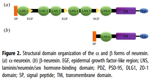
Critical periods
and CNTNAP2
Another factor to consider is the role
of developmental “critical periods,” when brain circuits involved in language
are particularly plastic. Disruption of CNTNAP2 has been linked to altered
neuronal connectivity and delayed circuit maturation, which may extend or shift
these windows of plasticity. If so, interventions that stabilize network
activity—such as T-type calcium channel modulation—might help enable more
effective language development during these periods. This could potentially
explain why some improvements, once initiated, continue even after treatment is
stopped.
This also raises the possibility that
timing may be critical. If language development depends on sensitive
developmental windows, and pathways involving CNTNAP2 alter the timing of
circuit maturation, then the age at which a treatment is given could determine
its effectiveness. Interventions such as T-type calcium channel modulation may
be more beneficial when applied during periods of higher neural plasticity, and
less effective once circuits have become more established. This could help
explain why any signal of benefit has been difficult to detect in routine
clinical use.
Conclusion
The study did not have a placebo
group. We know from many previous small studies that in most cases everyone
improved in autism studies, including those who were assigned the placebo.
Has Niesen identified a simple therapy
that will improve speech in autism?
If ethosuximide strongly improves
language, why has this not already been noticed?
Neurologists have used ethosuximide
for decades for autistic children with absence seizures, but it is not widely
recognized as a language-enhancing drug.
I expect there likely is a subgroup of
responders, but it will not be a silver bullet for all.
Ethosuximide is cheap, but it can have
some unusual side effects.
Zonisamide is more predictable than
Ethosuximide, but still can have problematic side effects, more so than drugs
like bumetanide or atorvastatin.
It may be the case that responders to
Ethosuximide do not need to take it permanently and that has to be factored
into the side effect assessment.
Any potential benefit is likely
limited to a specific subgroup, such as children with subtle absence seizures,
epileptiform activity, or abnormalities in calcium channel signaling. One
candidate subgroup involves mutations in the CNTNAP2 gene, which are associated
with language impairment, autism, and increased neuronal excitability.
Preclinical studies suggest that targeting T-type calcium channels in such
models can reduce hyperexcitability and improve behavioral features, raising
the possibility that drugs like ethosuximide may be more effective in
individuals with similar underlying biology.
CNTNAP2 is also regulated by TCF4, the
gene mutated in Pitt-Hopkins syndrome, a condition marked by profound speech
deficits. This points to overlapping biological pathways underlying language
impairment across different neurodevelopmental disorders and reinforces the
idea that identifying responders will be key to determining clinical value.
So, another idea for Pitt Hopkins
parents is to consider is Ethosuximide. Maybe the parents’ organisation should
contact Charles Niesen to make a small clinical trial, like the forthcoming Clemastine
one.