UA-45667900-1
Showing posts with label Memantine. Show all posts

Friday 13 January 2023

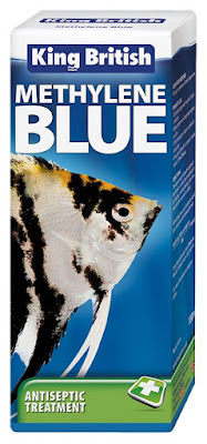
Methylene Blue - used for over a century in Psychiatry, also handy for your fish tank


According to the packaging:-

Effective against a range of fungal and bacterial infections

•          Increases the oxygen-carrying capacity of fish

•          Can be used as an antiseptic directly onto wounds

•          For use in tropical and cold water aquariums


Our reader Dragos recently let us all know about his success with very low doses of Methylene Blue (MB).  I think this came as a surprise to many, but actually there is nothing new about using this old pigment as a therapy in psychiatry.  Much is known about its modes of action.


What is Methylene Blue?

In 1876, German chemist Heinrich Caro synthesized methylene blue (MB) for the first time in history.  It was used as a dye for textiles. Around the same time, it was found that MB is capable of staining cells by binding to their structures, in addition, sometimes inactivating bacteria. This discovery prepared the way for biological or medical studies related to MB. Numerous scientists applied it to a variety of animal and bacterial studies, importantly Paul Ehrlich introduced it to humans in 1891 as an anti-malarial agent.

I was interested to see why it is used in aquariums, in particular the reference to increases the oxygen-carrying capacity of fish.

Methemoglobinemia (MetHb) is a rare blood disorder that affects how red blood cells deliver oxygen throughout your body.

A common way to treat  MetHb  in humans is to reduce methemoglobin levels using  Methylene blue (MB). Another common treatment, not surprisingly, is to give oxygen.

If you want to increase oxygen levels in the fish in your aquarium you put MB in the water.

More oxygen in your blood would improve exercise endurance meaning you would delay the point at which your mitochondria become unable to keep producing ATP efficiently.

I did some investigation and there is indeed a trend towards people using methyl blue to improve their sporting performance. It is mocked in some newspapers because it makes your tongue turn blue. It makes for good pictures on Instagram.     


The effect will be similar to those long distance cyclists who take beetroot juice, but the mechanism is different.

Be aware that just like beetroot may dye what comes out of your body bright red, MB may give you a hint of blue.


Improved Mitochondrial Function

One of the known effects of Methylene Blue (MB) is on the mitochondria.

In numerous papers it has been discussed how MB improves brain mitochondrial respiration.

In neurological disorders such as Alzheimer’s disease, traumatic brain injury, depression, stroke, Parkinson’s disease and some autism, mitochondria contribute to the disorder through decreased energy production and excessive production of reactive oxygen species (ROS).

This subject does get rather complex but in short methylene blue is able to perform alternative electron transport, bypassing parts of the electron transport chain.

In autism terms this means that some people diagnosed with a lack of Complex 1, 2, 3 or 4 in their mitochondria, might want to pay particular attention to how Methylene Blue might be helpful.

Improved mitochondrial function is another reason why sportsmen might want to use MB to enhance their performance.

As we have seen with other enhancing drugs like the Russian Meldonium, the US Diamox and the new US super ketone products, the military do end up using these products.  If you see a picture of a navy seal with a blue tongue you will know where it came from!


Methylene Blue inhibits Monoamine Oxidase (MAO)

MAOIs act by inhibiting the activity of monoamine oxidase, thus preventing the breakdown of monoamine neurotransmitters and thereby increasing their availability. There are two types of monoamine oxidase, MAO-A and MAO-B. MAO-A preferentially deaminates serotonin, melatonin, epinephrine, and norepinephrine. MAO-B preferentially deaminates phenethylamine and certain other trace amines; in contrast, MAO-A preferentially deaminates other trace amines, like tyramine, whereas dopamine is equally deaminated by both types.

Methyl blue is a reversible selective MAO-A inhibitor and so has antidepressant properties (it gives you more feel good serotonin). This interesting drug has several other pharmacological actions, including inhibition of nitric oxidase synthase (NOS), and guanylate cyclase and so its antidepressant properties should not be solely ascribed to inhibition of MAO-A. 

Inhibition of neuronal nitric oxide synthase and soluble guanylate cyclase prevents depression-like behaviour in rats exposed to chronic unpredictable mild stress

Beyond treating depression MAOIs (Monoamine oxidase inhibitors) have been found to be effective in the treatment of panic disorder, social phobia, mixed anxiety disorder and depression, bulimia, and post-traumatic stress disorder, as well as borderline personality disorder, and Obsessive Compulsive Disorder (OCD).

MAOIs appear to be particularly effective in the management of bipolar depression.

Methylene blue treatment for residual symptoms of bipolar disorder: randomised crossover study

Background: Residual symptoms and cognitive impairment are among important sources of disability in patients with bipolar disorder. Methylene blue could improve such symptoms because of its potential neuroprotective effects.

Aims: We conducted a double-blind crossover study of a low dose (15 mg, 'placebo') and an active dose (195 mg) of methylene blue in patients with bipolar disorder treated with lamotrigine.

Method: Thirty-seven participants were enrolled in a 6-month trial (trial registration: NCT00214877). The outcome measures included severity of depression, mania and anxiety, and cognitive functioning.

Results: The active dose of methylene blue significantly improved symptoms of depression both on the Montgomery-Åsberg Depression Rating Scale and Hamilton Rating Scale for Depression (P = 0.02 and 0.05 in last-observation-carried-forward analysis). It also reduced the symptoms of anxiety measured by the Hamilton Rating Scale for Anxiety (P = 0.02). The symptoms of mania remained low and stable throughout the study. The effects of methylene blue on cognitive symptoms were not significant. The medication was well tolerated with transient and mild side-effects.

Conclusions: Methylene blue used as an adjunctive medication improved residual symptoms of depression and anxiety in patients with bipolar disorder.


Methylene Blue activates oxidative stress response genes via Nrf2

One of the antioxidant effects of MB is activation of the redox switch Nrf2.  In the paper below it is also mentioned that MB has a beneficial against tau proteins. Amyloid and tau proteins clog up the brain in Alzheimer’s and as a result MB has been proposed as a therapy for dementia. 


Methylene blue upregulates Nrf2/ARE genes and prevents tau-related neurotoxicity

Methylene blue (MB, methylthioninium chloride) is a phenothiazine that crosses the blood brain barrier and acts as a redox cycler. Among its beneficial properties are its abilities to act as an antioxidant, to reduce tau protein aggregation and to improve energy metabolism. These actions are of particular interest for the treatment of neurodegenerative diseases with tau protein aggregates known as tauopathies. The present study examined the effects of MB in the P301S mouse model of tauopathy. Both 4 mg/kg MB (low dose) and 40 mg/kg MB (high dose) were administered in the diet ad libitum from 1 to 10 months of age. We assessed behavior, tau pathology, oxidative damage, inflammation and numbers of mitochondria. MB improved the behavioral abnormalities and reduced tau pathology, inflammation and oxidative damage in the P301S mice. These beneficial effects were associated with increased expression of genes regulated by NF-E2-related factor 2 (Nrf2)/antioxidant response element (ARE), which play an important role in antioxidant defenses, preventing protein aggregation, and reducing inflammation. The activation of Nrf2/ARE genes is neuroprotective in other transgenic mouse models of neurodegenerative diseases and it appears to be an important mediator of the neuroprotective effects of MB in P301S mice. Moreover, we used Nrf2 knock out fibroblasts to show that the upregulation of Nrf2/ARE genes by MB is Nrf2 dependent and not due to secondary effects of the compound. These findings provide further evidence that MB has important neuroprotective effects that may be beneficial in the treatment of human neurodegenerative diseases with tau pathology.


MB to treat inflammation and pain via sodium ion channels and iNOS

MB abates inflammation by suppressing nitric oxide production, and ultimately relieves pain in arthritis and colitis.  

MB suppresses the iNOS/NO-mediated inflammatory signaling by directly downregulating inducible NO synthase (iNOS).

Nitric oxide (NO) is a free radical which, in reactions with various molecules causes multiple biological effects, some good and some harmful.

It is produced by a reaction involving one of three enzymes iNOS, eNOS and nNOS.  i = inducible, n = neuronal and e = endothelial

iNOS is a major downstream mediator of inflammation.

eNOS is very helpful because it can widen blood vessels and so reduce blood pressure and increase blood flow.

nNOS is found in the brain and the peripheral nerve system where it has several important functions.  

MB may impede pain transmission by dampening neuronal excitability elicited by voltage-gated sodium channels (VGSCs).  You would then think that in people with seizures due to malfunctioning sodium channels, MB might be beneficial; for example Nav1.1 in Dravet syndrome. 

Methylene Blue Application to Lessen Pain: Its Analgesic Effect and Mechanism

Methylene blue (MB) is a cationic thiazine dye, widely used as a biological stain and chemical indicator. Growing evidence have revealed that MB functions to restore abnormal vasodilation and notably it is implicated even in pain relief. Physicians began to inject MB into degenerated disks to relieve pain in patients with chronic discogenic low back pain (CDLBP), and some of them achieved remarkable outcomes. For osteoarthritis and colitis, MB abates inflammation by suppressing nitric oxide production, and ultimately relieves pain. However, despite this clinical efficacy, MB has not attracted much public attention in terms of pain relief. Accordingly, this review focuses on how MB lessens pain, noting three major actions of this dye: anti-inflammation, sodium current reduction, and denervation. Moreover, we showed controversies over the efficacy of MB on CDLBP and raised also toxicity issues to look into the limitation of MB application. This analysis is the first attempt to illustrate its analgesic effects, which may offer a novel insight into MB as a pain-relief dye. 


Nicotinic acetylcholine receptors

The modulation of nicotinic acetylcholine receptors (nAChRs) has been suggested to play a role in the pathogenesis of various neurodegenerative diseases. 

MB acts as a non-competitive antagonist on α7 nAChRs.

Well known drugs that act in a similar way include the Alzheimer’s drug Memantine and Ketamine. Recall that intranasal Ketamine has been used in autism. 

Substances  with the opposite effect include nicotine, choline and of course

Amyloid beta, the marker of Alzheimer's disease.

Note that some people need to block α7 nAChRs and some people need to activate them. 

Methylene blue inhibits the function of α7-nicotinic acetylcholine receptors


FDA Drug Safety Communication: Serious CNS reactions possible when methylene blue is given to patients taking certain psychiatric medications

A list of the serotonergic psychiatric medications that can interact with methylene blue can be found here. 

  • Methylene blue can interact with serotonergic psychiatric medications and cause serious CNS toxicity.
  • In emergency situations requiring life-threatening or urgent treatment with methylene blue (as described above), the availability of alternative interventions should be considered and the benefit of methylene blue treatment should be weighed against the risk of serotonin toxicity. If methylene blue must be administered to a patient receiving a serotonergic drug, the serotonergic drug must be immediately stopped, and the patient should be closely monitored for emergent symptoms of CNS toxicity for two weeks (five weeks if fluoxetine [Prozac] was taken), or until 24 hours after the last dose of methylene blue, whichever comes first.
  • In non-emergency situations when non-urgent treatment with methylene blue is contemplated and planned, the serotonergic psychiatric medication should be stopped to allow its activity in the brain to dissipate. Most serotonergic psychiatric drugs should be stopped at least 2 weeks in advance of methylene blue treatment. Fluoxetine (Prozac), which has a longer half-life compared to similar drugs, should be stopped at least 5 weeks in advance.
  • Treatment with the serotonergic psychiatric medication may be resumed 24 hours after the last dose of methylene blue.
  • Serotonergic psychiatric medications should not be started in a patient receiving methylene blue. Wait until 24 hours after the last dose of methylene blue before starting the antidepressant.
  • Educate your patients to recognize the symptoms of serotonin toxicity or CNS toxicity and advise them to contact a healthcare professional immediately if they experience any symptoms while taking serotonergic psychiatric medications or methylene blue.


Conclusion 

Rather surprisingly, this therapy from the fish tank may have wide ranging effects on the autistic brain and in those with dementia, bipolar etc.

Possible benefits might include:

·        Improved production of ATP (energy) in the brain

·        Reduced oxidative stress in the brain

·        Reduced nitrosative stress

·        Reduced inflammation

·        Improved mood (due to increased serotonin)

·        Improved memory and cognitive function

·        Reduction in obsessive behaviors

In one of the papers, they comment that “methylene blue modulates functional connectivity in the human brain”.

It seems to work for Dragos.  You can also see that people on Reddit use it for issues like ADHD. 


Note the FDA warning:

Do not combine Methylene Blue with serotonergic psychiatric medications, because of the risk of serotonin syndrome (i.e., serotonin toxicity).


Wednesday 13 April 2022

Personalized/Precision Medicine for Sound Sensitivity in Autism, Bipolar and Schizophrenia?


Stop the Noise!


Conventional wisdom, even among enlightened neurologists like Manuel Casanova, is that you cannot medically treat the sensory issues that occur in neurological conditions like autism, bipolar and schizophrenia.

This blog is very much driven by the peer-reviewed literature, but very often seems to comes up with alternative interpretations to what the doctors will say.  Today is another of those days.

I do tell people that you can very easily get things 100% back to front when developing personalized/precision medicine.  The general idea was correct, but the effect was the exact opposite to what was hoped for.  This is not a failure; this is a learning experience.  Today we see that what works in schizophrenia is the exact opposite of what works in bipolar.  I do like to include schizophrenia and bipolar in my autism posts, because there is a big overlap between them and the broad umbrella of dysfunctions found in autism.

Sensory problems are very common in autism, bipolar and schizophrenia.

This post is mainly about issues with sound.  Vision is closely related. Smell, taste and texture may be less closely related. 

Sound/Hearing issues in autism 

Very often young children with autism do not respond to their name, or some other sounds; the natural first step is to check their hearing.  The majority of the time, their hearing turns out to be perfect.

As the child gets older and struggles with sounds like a baby crying, or a dog barking, parents may begin to feel their child’s hearing is too good!


The medical terms


Hyperacusis is a disorder in loudness perception and should mean you hear sounds too loudly.  The opposite term is hypoacusis and in the medical jargon it means you are going deaf, rather than having a volume perception problem

Tinnitus is hearing sounds that do not exist, but there are many possible causes.

Misophonia means hatred of sound, but those hated sounds are often very specific repeated human sounds like noisy eating, chewing, sniffing, coughing or machine-made sounds like a noisy clock ticking, or even a leaf blower.

There does appear to be a visual equivalent of sound Misophonia.

For some people, visual triggers can cause a similar reaction. This might happen if you see someone:

  • wagging their legs or feet (foot flapping)
  • rubbing their nose or picking at their finger nails
  • twirling their hair or pen
  •  chewing gum 

Some people suffer from a combination of sound disorders.  Many people with tinnitus also suffer from Misophonia. 

I think many people with autism are affected by a combination of Hyperacusis and Misophonia.

It seems that many people with Asperger’s suffer from hyperacusis, a substantial minority experience tinnitus. Almost all who suffer tinnitus also experience hyperacusis.

I think it might be hard to know if a person with severe autism and ID had tinnitus.


Tinnitus and hyperacusis in autism spectrum disorders with emphasis on high functioning individuals diagnosed with Asperger's Syndrome

Objectives: To evaluate the prevalence of tinnitus and hyperacusis in individuals with Asperger's Syndrome (AS).

Methods: A home-developed case-history survey and three item-weighted questionnaires: Tinnitus Reaction Questionnaire (TRQ), Tinnitus Handicap Inventory (THI), and the Hyperacusis Questionnaire (HQ) were employed. These tools categorize the subjective response to tinnitus and hyperacusis. The research tools were mailed to a mailing list of individuals with Asperger's Syndrome.

Results: A total of 55 subjects diagnosed with AS were included in the analysis (15.5% response rate). Sixty-nine percent of all respondents (38/55) reported hyperacusis with an average HQ score of 20.7. Furthermore, 35% (19/55) reported perceiving tinnitus with average scores of 27 for the TRQ and 23 for the THI. Thirty-one percent (17/55) reported both hyperacusis and tinnitus. The prevalence of hyperacusis in the AS respondents remained relatively constant across age groups.

Conclusions: Hyperacusis and tinnitus are more prevalent in the ASD population subgroup diagnosed with AS under DSM-IV criteria than in the general public. Hyperacusis also appears to be more prevalent in the AS population than in the ASD population at large. Future research is warranted to provide insight into the possible correlation between tinnitus and hyperacusis symptoms and the abnormal social interactions observed in this group.


All three terms are just observation diagnoses, they do not tell you what is the underlying biological cause.  In this blog we are interested in the underlying biology, because the goal is to find an effective treatment.

Hearing issues are common comorbities of well-known medical conditions; for example, people with type 1 diabetes may well suffer from tinnitus and hypoacusis.


Schematic block diagram of mechanisms that produce misophonia, hyperacusis, tinnitus, polycusis, and other false auditory percepts. Afferents from the cochlea, saccule, somesthetic pathways, and visceral sensory pathways contribute to processing in auditory lemniscal pathways. Modular thalamocortical processing is hypothesized to contribute (1) a common component to comorbid features of hyperacusis and tinnitus, (2) a component that produces unique features of tinnitus, and (3) component(s) for other false auditory perceptions. A parallel, interoceptive, and affective network produces the aversion, annoyance, fear, and pain-like features that may be associated with hyperacusis and misophonia

Source: https://www.ncbi.nlm.nih.gov/pmc/articles/PMC6453992/


 The research terms

The medical world is often rather short of enough descriptive words, just think about all those people with totally different biological conditions all being diagnosed with “autism”.

A really useful term you will find in the research is sensory gating.


Sensory gating is a process by which irrelevant stimuli are separated from meaningful ones.  Imagine the boy with Asperger’s sitting in a private room taking his important exams.  He is alone with the invigilator and maybe a clock on the wall.  The clock might be making a ticking sound or the invigilator might be chewing gum.  All this clever boy has to do is to concentrate on the exam and show how smart he is.  The noisy clock, or the chewing sound, should be irrelevant, but instead the boy cannot filter out these sounds and ignore them.

I had exactly this case put to me at an autism conference by a concerned Grandfather, whose clever grandson failed his important exams.

You can actually measure sensory gating using headphones to provide the annoying repetitive sound and an EEG to measure how the person’s brain responds.  The first sound should trigger the brain’s response, but when the sound keeps repeating the response should fade away.  The person has learned to filter out the annoying but irrelevant sound.

Imagine you are in a storm and the rain is beating down on a glass roof or windows.  The first sound alerts you to the storm.  Did you leave the upstairs window open? Perhaps you were drying something outside?  You might have to take some urgent action, so you want an alarm bell to go off in your head.  Panic over, you can then just ignore the sound of the rain and before you know it the storm is over.

There are different types of sensory gating, the most well studied is called P50.

People with schizophrenia often have deficits in gating the neuronal response of the P50 wave, which is why P50 is the most widespread method of diagnosis. The test is conducted through having the patients hear two uniform sounds with an interval of 500 milliseconds. While the patients are hearing the sound, an EEG cap is used to measure the brain activity in response to those sounds. A normal subject shows a decrease in brain activity while hearing a second sound, while a subject showing equal brain activity to the first sound has impaired sensory gating.

Impaired P50 sensory gating is very common in schizophrenia, also occurs in autism bipolar and even dementia.

There can also be Impaired gating of N100 and P200.  The actual definition of these terms gets complicated and you do not have to go into this level of detail unless you are really interested


What is N100 event-related potential? 

The N100 is a negative waveform that peaks at approximately 100 milliseconds after stimulus presentation. Its amplitude is measured using electroencephalography (EEG) and may be dysfunctional in people with schizophrenia who show an inability to “gate” or inhibit irrelevant sensory information, ultimately leading to conscious information overload. To test this, paired auditory clicks are presented, separated by a short interval, usually of 0.5 seconds. The first click initiates or conditions the inhibition, while the second (test) click indexes the strength of the inhibition. An absence of a reduced response to the second stimulus is interpreted as a failure of inhibitory mechanisms, postulated to represent a defect in sensory gating.


What is the evidence for N100 event-related potential? 

Moderate to high quality evidence finds a medium-sized reduction in N100 amplitude to the first stimulus, but not to the second stimulus. Review authors suggests this reflects a deficit in processing of auditory salience rather than in inhibition.


P50-N100-P200 sensory gating deficits in adolescents and young adults with autism spectrum disorders


Highlights 

·        In the paired-click paradigm, ASD individuals displayed a significant N100 gating deficit.

·        N100 gating deficit was associated with symptom severity of sensory sensitivity.

·        P50 and P200 in ASD did not deviate from the typically developing controls.

·        P50 and P200 were associated with social deficits and attention switching difficulty in ASD.

 We found that compared to TDC, ASD participants had significant N100 suppression deficits reflected by a larger N100 S2 amplitude, smaller N100 ratio of S2 over S1, and the difference between the two amplitudes. N100 S2 amplitude was significantly associated with sensory sensitivity independent of the diagnosis. Although there was no group difference in P50 suppression, S1 amplitude was negatively associated with social deficits in ASD. P200 gating parameters were correlated with attention switching difficulty. Our findings suggest N100 gating deficit in adolescents and young adults with ASD. The relationships between P50 S1 and social deficits and between N100 S2 and sensory sensitivity warrant further investigation.


Expanding our understanding of sensory gating in children with autism spectrum disorders


Highlights


·        Children with autism showed significantly reduced gating at P50, N1, and P2 event-related potential components.

·        Children with autism show reduced orientation to auditory stimuli compared to typically-developing children.

·        Time-frequency analysis show reduced neural synchronization of stimuli in children with autism.

Abstract

Objective

This study examined sensory gating in children with autism spectrum disorders (ASD). Gating is usually examined at the P50 component and rarely at mid- and late-latency components.

Methods

Electroencephalography data were recorded during a paired-click paradigm, from 18 children with ASD (5–12 years), and 18 typically-developing (TD) children. Gating was assessed at the P50, N1, P2, and N2 event-related potential components. Parents of all participants completed the Short Sensory Profile (SSP).

Results

TD children showed gating at all components while children with ASD showed gating only at P2 and N2. Compared to TD children, the ASD group showed significantly reduced gating at P50, N1, and P2. No group differences were found at N2, suggesting typical N2 gating in the ASD group. Time-frequency analyses showed reduced orientation and neural synchronization of auditory stimuli. P50 and N1 gating significantly correlated with the SSP.

Conclusion

Although children with ASD have impaired early orientation and filtering of auditory stimuli, they exhibited gating at P2 and N2 components suggesting use of different gating mechanisms compared to TD children. Sensory deficits in ASD may relate to gating.

Significance

The data provide novel evidence for impaired neural orientation, filtering, and synchronization in children with ASD.


Normal P50 Gating in Children with Autism, Yet Attenuated P50 Amplitude in the Asperger Subcategory 

Autism spectrum disorders (ASD) and schizophrenia are separate disorders, but there is evidence of conversion or comorbid overlap. The objective of this paper was to explore whether deficits in sensory gating, as seen in some schizophrenia patients, can also be found in a group of ASD children compared to neurotypically developed children. An additional aim was to investigate the possibility of subdividing our ASD sample based on these gating deficits. In a case–control design, we assessed gating of the P50 and N100 amplitude in 31 ASD children and 39 healthy matched controls (8–12 years) and screened for differences between groups and within the ASD group. We did not find disturbances in auditory P50 and N100 filtering in the group of ASD children as a whole, nor did we find abnormal P50 and N100 amplitudes. However, the P50 amplitude to the conditioning stimulus was significantly reduced in the Asperger subgroup compared to healthy controls. In contrast to what is usually reported for patients with schizophrenia, we found no evidence for sensory gating deficits in our group of ASD children taken as a whole. However, reduced P50 amplitude to conditioning stimuli was found in the Asperger group, which is similar to what has been described in some studies in schizophrenia patients. There was a positive correlation between the P50 amplitude of the conditioning stimuli and anxiety score in the pervasive developmental disorder not otherwise specified group, which indicates a relation between anxiety and sensory registration in this group


Treatments for sensory gating

We know that in schizophrenia impaired P50 gating is associated with alpha 7 nicotinic acetylcholine receptor (α7 nAChR) dysfunction and shown to be improved with nicotine and other α7 nAChR agonists.

Other α7 nAChR agonists include:-

·        Acetylcholine

·        Choline

·        Nicotine

·        Tropisetron


Galantamine is a positive allosteric modulator (PAM) of nAChRs


Why do people with schizophrenia love to smoke?


A truly remarkable observation is that smoking improves sensory gating in schizophrenia, but it has the opposite effect on people with bipolar.


Smoking as a Common Modulator of Sensory Gating and Reward Learning in Individuals with Psychotic Disorders


Motivational and perceptual disturbances co-occur in psychosis and have been linked to aberrations in reward learning and sensory gating, respectively. Although traditionally studied independently, when viewed through a predictive coding framework, these processes can both be linked to dysfunction in striatal dopaminergic prediction error signaling. This study examined whether reward learning and sensory gating are correlated in individuals with psychotic disorders, and whether nicotine—a psychostimulant that amplifies phasic striatal dopamine firing—is a common modulator of these two processes. We recruited 183 patients with psychotic disorders (79 schizophrenia, 104 psychotic bipolar disorder) and 129 controls and assessed reward learning (behavioral probabilistic reward task), sensory gating (P50 event-related potential), and smoking history. Reward learning and sensory gating were correlated across the sample. Smoking influenced reward learning and sensory gating in both patient groups; however, the effects were in opposite directions. Specifically, smoking was associated with improved performance in individuals with schizophrenia but impaired performance in individuals with psychotic bipolar disorder. These findings suggest that reward learning and sensory gating are linked and modulated by smoking. However, disorder-specific associations with smoking suggest that nicotine may expose pathophysiological differences in the architecture and function of prediction error circuitry in these overlapping yet distinct psychotic disorders.


When you look up P50 gating and also Misophonia in the clinical trials database, you get some Mickey Mouse behavioral treatments for misophonia.

For p50 gating you a decent list of drugs trialed in schizophrenia. 


My earlier posts on this subject:-


Sensory Gating in Autism, Particularly Asperger's


Cognitive Loss/Impaired Sensory Gating from HCN Channels - Recovered by PDE4 Inhibition or an α2A Receptor Agonist


"I did wonder how nicotine fits in, since in earlier post we saw that α7 nAChR agonists, like nicotine, improve sensory gating and indeed that people with schizophrenia tend to be smokers. It turns out that nicotine is also an HCN channel blocker. For a change, everything seems to fit nicely together. There are different ways to block HCN channels, some of which are indirect. One common ADHD drug, Guanfacine, keeps these channels closed, but in a surprising way."


Acute administration of Roflumilast enhances sensory gating in healthy young humans in a randomized trial. 


Abstract


INTRODUCTION:

Sensory gating is a process involved in early information processing which prevents overstimulation of higher cortical areas by filtering sensory information. Research has shown that the process of sensory gating is disrupted in patients suffering from clinical disorders including attention deficit hyper activity disorder, schizophrenia, and Alzheimer's disease. Phosphodiesterase (PDE) inhibitors have received an increased interest as a tool to improve cognitive performance in both animals and man, including sensory gating.

METHODS:

The current study investigated the effects of the PDE4 inhibitor Roflumilast in a sensory gating paradigm in 20 healthy young human volunteers (age range 18-30 years). We applied a placebo-controlled randomized cross-over design and tested three doses (100, 300, 1000 μg).

RESULTS:

Results show that Roflumilast improves sensory gating in healthy young human volunteers only at the 100-μg dose. The effective dose of 100 μg is five times lower than the clinically approved dose for the treatment of acute exacerbations in chronic obstructive pulmonary disease (COPD). No side-effects, such as nausea and emesis, were observed at this dose. This means Roflumilast shows a beneficial effect on gating at a dose that had no adverse effects reported following single-dose administration in the present study.

CONCLUSION:

The PDE4 inhibitor Roflumilast has a favourable side-effect profile at a cognitively effective dose and could be considered as a treatment in disorders affected by disrupted sensory gating.


Be wary of antipsychotics!!

 Now we see again that α2A Receptor agonists like guanfacine and clonidine will improve sensory gating. We should not be surprised that drugs with the opposite effect (antagonists) will make sensory gating worse.


α2A Receptor Antagonists

·         Idazoxan

·         1-PP (active metabolite of buspirone and gepirone, anti-anxiety drugs)

·         Asenapine

·         BRL-44408

·         Clozapine , an anti-psychotic drugs used in schizophrenia

·         Lurasidone an anti-psychotic drugs used in schizophrenia and in bipolar

·         Mianserin, an anti-depressant

·         Mirtazapine, an anti-depressant

·         Paliperidone an anti-psychotic drugs used in schizophrenia

·         Risperidone, an anti-psychotic drugs used in schizophrenia and autism

·         Yohimbine


Treatment for Hyperacusis

If you look up treatments and trials for hyperacusis (sound sensitivity) you see a list of cognitive behavioral therapies.

These are not nonsense. We used something similar to deal with Monty’s extreme aversion to crying babies when he was young.  Now when he hears a baby crying, he laughs.

But really, science has much more to offer than behavioral therapy.

I did write many years ago about hypokalemic sensory overload and its big brother hypokalemic periodic paralysis (HypoPP).  In both conditions it seems that low levels of potassium cause some pretty severe reactions.  Both conditions respond rapidly to an oral potassium supplement.

Though rare, we know that HypoPP is caused by a dysfunction in the ion channels Nav1.4 and/or Cav1.1.

For decades one of the treatments for HypoPP has been a diuretic called Diamox/Acetazolamide.  Other treatments include raising potassium levels using supplements, or potassium sparing diuretics.


Way back in 2013, I defined a new term, in the post below:-


 Hypokalemic Autistic Sensory Overload


I showed an oral potassium supplement reduced sound sensitivity within 20 minutes, with a simple experiment anyone can do at home. 

Some people do find long term sensory relief just from the use of an oral potassium supplement once a day.  In my son’s case the affect does not last very long.


Therapies for hypokalemic sensory overload might be:-


·        A potassium supplement

·        A potassium sparing diuretic

·        Possibly Diamox/ Acetazolamide

·        Very likely, intra-nasal Desmopressin, this lower sodium levels and so will have the opposite impact on potassium levels

·        Ponstan, the NSAID that affects numerous potassium ion channels


In some people it appears that Humira, a long-acting TNF-alpha inhibitor, resolves visual and sound sensitivity.  I think this resolves a mixture of hyperacusis and Misophonia and the visual sensory equivalents.


Tinnitus

Tinnitus is an extremely common, but is generally regarded as something you just have to get used to; there are no approved drug therapies.

All kinds of things can lead to tinnitus. A head injury can lead to tinnitus, exposure to a loud sound is a common cause, but there is even drug-induced tinnitus. Tinnitus is a common comorbidity of diabetes.

There is gradual onset tinnitus and acute onset tinnitus.

Tinnitus is more likely to occur the older you get and often gets worse over time.

Clearly there are many sub-types of tinnitus and inevitably there will need to be multiple different therapies


Full graphic is available at fnins-13-00802-g004.jpg (4660×2924) (frontiersin.org)


The paper below is very comprehensive: 

Why Is There No Cure for Tinnitus? 

Tinnitus is unusual for such a common symptom in that there are few treatment options and those that are available are aimed at reducing the impact rather than specifically addressing the tinnitus percept. In particular, there is no drug recommended specifically for the management of tinnitus. Whilst some of the currently available interventions are effective at improving quality of life and reducing tinnitus-associated psychological distress, most show little if any effect on the primary symptom of subjective tinnitus loudness. Studies of the delivery of tinnitus services have demonstrated considerable end-user dissatisfaction and a marked disconnect between the aims of healthcare providers and those of tinnitus patients: patients want their tinnitus loudness reduced and would prefer a pharmacological solution over other modalities. Several studies have shown that tinnitus confers a significant financial burden on healthcare systems and an even greater economic impact on society as a whole. Market research has demonstrated a strong commercial opportunity for an effective pharmacological treatment for tinnitus, but the amount of tinnitus research and financial investment is small compared to other chronic health conditions. There is no single reason for this situation, but rather a series of impediments: tinnitus prevalence is unclear with published figures varying from 5.1 to 42.7%; there is a lack of a clear tinnitus definition and there are multiple subtypes of tinnitus, potentially requiring different treatments; there is a dearth of biomarkers and objective measures for tinnitus; treatment research is associated with a very large placebo effect; the pathophysiology of tinnitus is unclear; animal models are available but research in animals frequently fails to correlate with human studies; there is no clear definition of what constitutes meaningful change or “cure”; the pharmaceutical industry cannot see a clear pathway to distribute their products as many tinnitus clinicians are non-prescribing audiologists. To try and clarify this situation, highlight important areas for research and prevent wasteful duplication of effort, the British Tinnitus Association (BTA) has developed a Map of Tinnitus. This is a repository of evidence-based tinnitus knowledge, designed to be free to access, intuitive, easy to use, adaptable and expandable.


The next paper makes the key point that to treat tinnitus you need precision (personalized) medicine and apply the neuroscience.


Towards a Mechanistic-Driven Precision Medicine Approach for Tinnitus 

In this position review, we propose to establish a path for replacing the empirical classification of tinnitus with a taxonomy from precision medicine. The goal of a classification system is to understand the inherent heterogeneity of individuals experiencing and suffering from tinnitus and to identify what differentiates potential subgroups. Identification of different patient subgroups with distinct audiological, psychophysical, and neurophysiological characteristics will facilitate the management of patients with tinnitus as well as the design and execution of drug development and clinical trials, which, for the most part, have not yielded conclusive results. An alternative outcome of a precision medicine approach in tinnitus would be that additional mechanistic phenotyping might not lead to the identification of distinct drivers in each individual, but instead, it might reveal that each individual may display a quantitative blend of causal factors. Therefore, a precision medicine approach towards identifying these causal factors might not lead to subtyping these patients but may instead highlight causal pathways that can be manipulated for therapeutic gain. These two outcomes are not mutually exclusive, and no matter what the final outcome is, a mechanistic-driven precision medicine approach is a win-win approach for advancing tinnitus research and treatment. Although there are several controversies and inconsistencies in the tinnitus field, which will not be discussed here, we will give a few examples, as to how the field can move forward by exploring the major neurophysiological tinnitus models, mostly by taking advantage of the common features supported by all of the models. Our position stems from the central concept that, as a field, we can and must do more to bring studies of mechanisms into the realm of neuroscience.


I did have a quick look the clinical trials website to see if there have been any interesting trials that did show some benefit. 

I noted the following drugs: 

Lidocaine

Lidocaine, the anesthetic that targets sodium ion channels.  Careful titration allows for a high degree of selectivity in the blockage of sensory neurons.  This looks like a good idea. Originally, they played with intravenous delivery, but then moved no to transdermal.


Transdermal lidocaine as treatment for chronic subjective tinnitus: A Pilot Study

In this preliminary study, 5% transdermal lidocaine appears to be a potential treatment for chronic subjective tinnitus. The majority of subjects who completed 1 month of treatment had clinically significantly improved tinnitus. These findings are confounded however by the small sample size and significant drop out rate.


Clonazepam 

Clonazepam is a benzodiazepine drug that activates GABAa receptors.  The trials are a bit mixed and one showed it only worked when given together with Deanxit. Deanxit is a combination of Flupentixol, an antipsychotic, and melitracen an tricyclic antidepressant.

These look like bad options which will end up causing new problems over time. 

Clonazepam Quiets tinnitus: a randomised crossover study with Ginkgo Biloba

Conclusion Clonazepam is effective in treating tinnitus; G biloba is ineffective.


Administration of the combination clonazepam-Deanxit as treatment for tinnitus

Results: Significant tinnitus reduction was seen after intake of the combination clonazepam-Deanxit, whereas no differences in tinnitus could be demonstrated after the administration of clonazepam-placebo. This was true for all patients according to the following parameters: time patients are annoyed by the tinnitus (p = 0.026) and the visual analogue scale for tinnitus annoyance (p = 0.024).

 Conclusion: Although tinnitus reduction was recorded as modest, this article provides valuable data demonstrating a placebo-controlled tinnitus reduction after clonazepam and Deanxit intake.


Oxytocin

There already is a lot in the blog about oxytocin and I was surprised anyone had trialed it for tinnitus, but they did and it seems to provide a benefit.  As regular readers of this blog know, there looks to be a better way to deliver oxytocin to the brain than intra-nasal. We saw how a specific gut bacteria has the same effect (Biogaia Protectis). 

TinnitusTreatment with Oxytocin: A Pilot Study

Conclusion

These preliminary studies demonstrated that oxytocin may represent a helpful tool for treating tinnitus and further larger controlled studies are warranted.


Acamprosate

Acamprosate is used to treat alcoholics.

 “An inhibition of the GABA-B system is believed to cause indirect enhancement of GABAA receptors.[17] The effects on the NMDA complex are dose-dependent; the product appears to enhance receptor activation at low concentrations, while inhibiting it when consumed in higher amounts, which counters the excessive activation of NMDA receptors in the context of alcohol withdrawal”  

Impact of Acamprosate on Chronic Tinnitus: A Randomized-Controlled Trial 

Objectives: Tinnitus is a common and distressing otologic symptom, with various probable pathophysiologic mechanisms, such as an imbalance between excitatory and inhibitory mechanisms. Acamprosate, generally used to treat alcoholism, is a glutaminergic antagonist and GABA agonist suggested for treating tinnitus. Thus, we aimed to evaluate the efficacy and safety of acamprosate in the treatment of tinnitus.

Conclusions: The study results indicated a subjective relief of tinnitus as well as some degree of the electrophysiological improvement at the level of the cochlear and the distal portion of the auditory nerve among the subjects who received the acamprosate.


Magnesium

Magnesium supplementation, being cheap and OTC, is a common therapy for tinnitus.  It does seem to provide a benefit for some. 

Phase 2 study examining magnesium-dependent tinnitus

Conclusion: The results suggest that magnesium may have a beneficial effect on perception of tinnitus-related handicap when scored with the THI.


Neramexane

Neramexane is interesting because it is closely related to Memantine/Namenda, which was widely used in autism, but failed in its large clinical trial.  Memantine is seen as an NMDA receptor antagonist/blocker, but it also blocks  nicotinic acetylcholine receptors (nAChRs) which play a role in Alzheimer’s and sensory gating (Misophonia). Memantine also affects serotonin and dopamine receptors.

 Neramexane is a new drug being developed for Alzheimer’s and as a pain killer. 

A randomized, double-blind, placebo-controlled clinical trial to evaluate the efficacy and safety of neramexane in patients with moderate to severe subjective tinnitus


Neramexane is a new substance that exhibits antagonistic properties at α9α10 cholinergic nicotinic receptors and N-methyl-D-aspartate receptors, suggesting potential efficacy in the treatment of tinnitus.


Conclusions

This study demonstrated the safety and tolerability of neramexane treatment in patients with moderate to severe tinnitus. The primary efficacy variable showed a trend towards improvement of tinnitus suffering in the medium- and high-dose neramexane groups. This finding is in line with consistent beneficial effects observed in secondary assessment variables. These results allow appropriate dose selection for further studies.


Mirtazapine 

Mirtazapine is yet another drug that has been covered in this blog.  It is a very cheap anti-histamine / anti-depressant.

We saw in this blog that the effect is highly dose dependent.  It affects very many receptors and the overall effect depends on dosage. The antidepressant effect is at the dose of 15+mg.  In this person with tinnitus, they used 7.5mg. For some conditions the dose goes up to 60mg a day.

At very low dosages mirtazapine is a potent H1 anti-histamine and makes you very drowsy

One parent noted that low dose Mirtazapine had a highly beneficial effect in their child with autism.


Tinnitus Treatment With Mirtazapine

Auditory pathways are modulated by various neurotransmitters such as serotonin responsible for sound detection, location, and interpretation. The neurotransmitter gamma amino butyric acid (GABA) is inhibitory in the auditory system. Given that there is preferential innervation of the GABAergic neurons in the inferior colliculus by serotonergic neurons, it may be plausible then that antidepressant drugs, by increasing the availability of serotonin and thereby increasing GABAergic activity, provide relief from the symptoms of tinnitus.5 This report shows that mirtazapine may have a beneficial effect in the subgroup of patients suffering from tinnitus but exact mechanism is difficult to put forward.


Conclusion 

I think we are absolutely spoilt for choice.

So many possible therapies, each one effective in some cases.

The key is precision medicine, personalized to the individual case in question.  This approach was also proposed in the recent paper on Tinnitus, only without telling us what to actually do!

In my son, now 18 with what we can call treated severe autism, the clear winner so far is Ponstan (Mefenamic Acid).  Diclofen, a very common Fenamate class drug, does share the same effect, but to a lesser extent. 

Fenamates (Diclofenac, Ponstan etc): certainly for Alzheimer’s, maybe some Epilepsy, but Autism? I’m Impressed!


Low dose Roflumilast, the P50 sensory gating therapy (that is more for Aspies) has no sensory effect at all. It is the same dose as that proposed in the research to raise IQ.

The intranasal Desmopressin mentioned by one reader is another good choice to consider, but you may need to supplement sodium.  I think if you get a short term benefit from a 500mg potassium supplement, this is worth a try.

For Aspies low dose Roflumilast everyday looks worth a try, while Humira every 2 months look interesting, but it will be hard to get and is pricey.

For people with Schizophrenia, they could look at tobacco alternatives, which would include low-dose Roflumilast.

People with Bipolar might want to look at Mirtazapine – the opposite of nicotine and which also helps some cases of tinnitus.

For tinnitus I thought oxytocin looked a very safe option.  You have intranasal, or my preference the gut bacteria probiotic that stimulates oxytocin release in the brain.

Magnesium is a safe bet for tinnitus.  Transdermal lidocaine makes sense, but is a bit more daring.  Memantine might be worth a shot, if nothing else helps.

You can also increase sound and visual sensitivity. Low dose DMF (dimethyl fumarate) increases sound sensitivity and the TRH super-agonist Ceredist increases visual sensitivity.  For most people with autism, you likely do not need either effect.